
Gynaecology
- Overview
- Equipment
- Our Doctors
- Patient Education Videos
Moreland Surgery Center offers a full array of gynecological surgical procedures to help women maintain their gynecological health. Some of the more common procedures performed at our center include:
Hysteroscopy
Hysteroscopy provides a way for your physician to look inside your uterus. A hysteroscope is a thin, telescope-like instrument that is inserted into the uterus through the vagina and cervix. This tool often helps a physician diagnose or treat a uterine problem. Hysteroscopy is minor surgery which is performed either in your physician's office or in a operating room setting. It can be performed with local, regional, or general anesthesia--sometimes no anesthesia is needed. There is little risk involved with this procedure for most women. Hysteroscopy may be either diagnostic or operative.
Diagnostic hysteroscopy is used to diagnose some uterine abnormalities, and may also be used to confirm the results of other tests such as hysterosalpingography (HSG). Other instruments or techniques, such as dilation and curettage (D&C) and laparoscopy, are sometimes used in conjunction with the hysteroscopy. Diagnostic hysteroscopy can be used to diagnose certain conditions such as abnormal uterine bleeding, infertility, repeated miscarriages, adhesions, fibroid tumors, polyps, or to locate displaced intrauterine devices (IUDs).
An operative hysterocopy may be used, instead of open abdominal surgery, to both diagnose and treat certain conditions such as uterine adhesions, septums, or fibroids which can often be removed through the hysteroscope.
Endometrial Ablation
The hysteroscope is sometimes used with other instruments such as the resectoscope to treat some cases of abnormal bleeding; however after this procedure, known as endometrial ablation, women can no longer have children so it is not an option for women who wish to have future pregnancies. Endometrial ablation is a procedure which destroys the lining of the uterus. The resectoscope is a telescope-like instrument with a wire loop, a rollerball, or a roller cylinder tip at the end. Electric current at the end of the tip is used to destroy the uterine lining. This procedure is usually performed in an outpatient setting.
Hysteroscopic Myomectomy
Submucous and intracavitary myomas can often be removed through the cervix using an instrument called a resectoscope. The resectoscope is a special type of hysteroscope with a built in wire loop that uses high-frequency electrical energy to cut or coagulate tissue. It was developed for surgery of the bladder and the male prostate over fifty years ago to allow surgery inside an organ without having to make an incision, and has made hysteroscopic myomectomy possible.
Just as with a standard hysteroscope, the resectoscope is inserted through the cervix. Because it goes through the cervix, it is not necessary to make an incision. Procedures using the resectoscope are done in an operating room setting. This can at times be done under local anesthesia, but most women prefer to be completely asleep with general anesthesia.
The resectoscope is far more efficient at removing tissue (such as fibroids or large polyps) than conventional instruments. Because of this, the resectoscope should be used only by physicians who have training in its use and who use it on a regular basis.
Dilation & Curettage
Often used to diagnose or treat abnormal uterine bleeding, the D&C is one of the most common surgical procedures performed on women. Dilation and Curettage also provides important information about whether uterine cancer is present. A D&C may be required to diagnosed and/or treat a problem such as heavy or prolonged menstruation, as well as unexplained bleeding between periods. Where your D&C takes place depends on individual factors about your health. It can be performed in an operating room setting using general anesthesia or in your doctor's office using a local anesthetic. An injection around the cervix will minimize pain or discomfort from the procedure and produce numbness in the area.
The doctor completely inspects the pelvic reproductive organs for any abnormal changes. Next, a speculum is inserted into the vagina to open the walls so the doctor can see the cervix. A clamp-like instrument holds the cervix in place. The cervix is dialated with a series of tapered rods of increasing widths which are inserted into the cervical opening (the OS). A curette is passed through the uterus and used to scrape the uterine walls. This loosens pieces of the lining which are removed and sent to a lab for microscopic examination. Another method of obtaining a sample of the uterine lining is by applying suction through a narrow tube.
LEEP Procedure
If your doctor has told you that you need to have a LEEP procedure, it's because your annual Pap smear indicated the presence of abnormal cervical cells, or cervical dysplasia. The loop electrosurgical excision procedure, or LEEP procedure, is one of several procedures your doctor has available to help diagnose and treat abnormal cervical cells. Other procedures your doctor may want you to have during the LEEP procedure include, a colposcopy and / or a cone biopsy.
If your doctor has told you that you need to have a LEEP procedure, it's because your annual Pap smear indicated the presence of abnormal cervical cells, or cervical dysplasia. The loop electrosurgical excision procedure, or LEEP procedure, is one of several procedures your doctor has available to help diagnose and treat abnormal cervical cells. Other procedures your doctor may want you to have during the LEEP procedure include, a colposcopy and / or a cone biopsy.
An electrosurgical dispersive pad will be placed on your thigh. The pad is a gel-covered adhesive electrode which provides a safe return path for the electrosurgical current. A single-use, disposable loop electrode will be attached to the generator hand piece by your physician. Your cervix will be prepared with acetic acid and iodine solutions that enable your physician to more easily see the extent of the abnormal area. Next a local anesthetic will be injected into the cervix; the electroloop will be generated and the wire loop will pass through the surface of your cervix.
After the lesion is removed your physician will use a ball electrode to stop any bleeding that occurs; he or she may also use a topical solution to prevent further bleeding. You can usually go home soon after the procedure.
Pelvic Laparoscopy
Laparoscopy is usually performed under general anesthesia; however it can be performed with other types of anesthesia that permit the patient to remain awake. The typical pelvic laparoscopy involves a small (1/2" to 3/4") incision in the belly button or lower abdomen. The abdominal cavity is filled with carbon dioxide. Carbon dioxide causes the abdomen to swell, which lifts the abdominal wall away from the internal organs. That way, the doctor has more room to work.
Next, a laparoscope (a one-half inch fiber-optic rod with a light source and video camera) is inserted through the belly button. The video camera permits the surgeon to see inside the abdominal area on video monitors located in the operating room.
Depending on the reason for the laparoscopy, such as tubal ligation, the physician may perform surgery through the laparoscope by inserting various instruments into the laparoscope while using the video monitor as a guide. The video camera also allows the surgeon to take pictures of any problem areas he discovers.
Click here for more information
Novasure
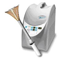
The NovaSure procedure is a quick, safe, simple, one-time endometrial ablation treatment. This minimally invasive procedure controls heavy bleeding by using energy to remove the lining of the uterus. The average treatment time is about 90 seconds 1, and only needs to be performed once to lighten or stop your periods. No pretreatment drugs are required and NovaSure can be performed in the hospital or in your doctor's office.
Without the side effects of hormones or the risks of hysterectomy, NovaSure has a quick recovery time so you can get back to your life sooner. Most women experience no pain after the procedure, and can return to work and regular activities the next day.